
Strive to Improve Medical Conditions, Reduce Health Care Costs
Strive to Improve Medical Conditions, Reduce Health Care Costs

Full support perspective, gastrointestinal spot film, GI (barium meal, barium enema), orthopedic photography, pediatrics photography, chest, urinary system angiography, peripheral angiography operations, gynecological Photography (HSG) and many other checks, the real machine can realize all these function.
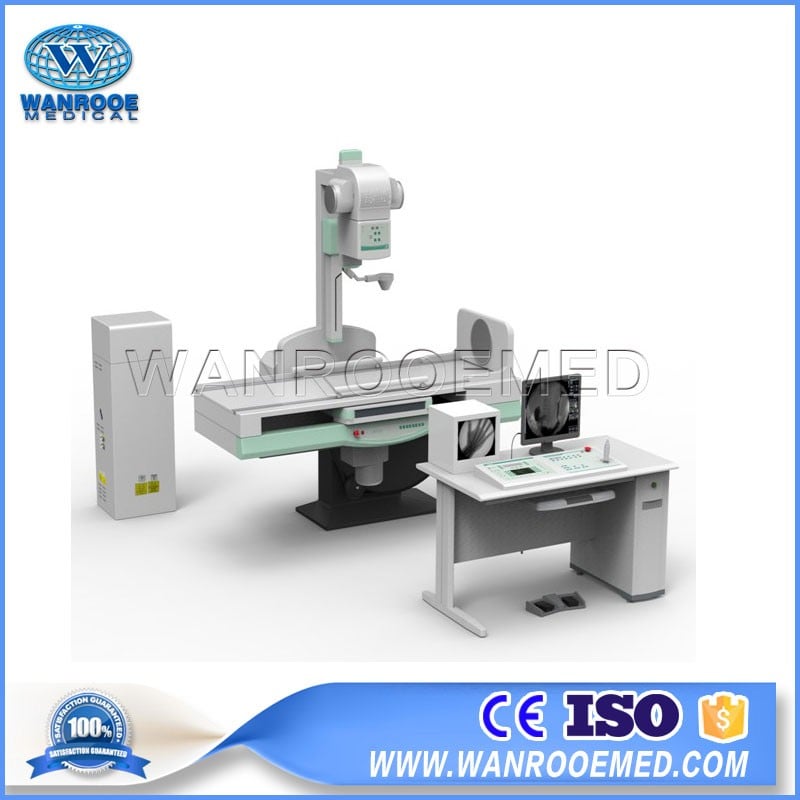
Main parts for the Machine
1.Table(It can up and down by manual)
2. High pressure system
1)High Frequency High Voltage X ray generator : CPI Brand made in Canada
2)Max Inverter Frequency : 80 kw, 200 KHZ
3)Constant DC output, to make sure high quality monochromatic X ray can be obtained,so that can eliminate the soft ray effect the image completely
4)Ultra high frequency high voltage generator, global leading high technology,X ray dose output precision, green technology, reduce emission lines
3. Equipped with high performance, large capacity of the X-ray tube
Use 0.6/1.2 mm 2 double focus, 300 khu large-capacity high-speed X-ray tube, the clinical examination for high intensity for a long time
Imported small focus, large-capacity high-speed X-ray tube components, suitable for long time and high strength of clinical examination
4. Image chain systems
Imported ultra metal screen image intensifier, 1 mage pixels.
Import Integrated Image Intensifier, three vision, 0.47 mega pixels
1)High quality metal screen image intensifier, cooperated with small focus X-ray tube and digital CCD camera, so that high-definition digital image can be obtained
2)Combination of CCD camera and digital image processor to realize the low noise, rich contrast perspective images, edge enhancement filter device make the image more clear and sharp edges.
3)High contrast pulse strengthens fluroscopy function , to ensure the patient can get clear diagnostic imaging even if the x ray does is only 10% of original one
5.Work station
Integrated combined operation handle, can easily control the bed body, image system and the movement of rotary pedal , spot film;
1)The table body can +90 degrees to 0 to -25 degrees rotate.
2)Humanized design of diagnosis bedside switch operation, can control the table body and imaging system movement,so that the close table inspection is convenient and easy operation;
3)Spot film device and imaging system movement range more than 720 mm
4)Adopts the operation of the machine move, but the patient don't need move, can easily finish from the throat, esophagus to the lower abdomen of a series of inspections;
5)Cassette trolley can test cassette size by itself, a key can completely finish the insert piece, save space, convenient and fast;
6. Table operation
Electric control of the infinite place 360 degrees rotation of the pedals
7. Image processing system
1 mega pixels, with digital workstation
Integration configuration: graphic workstation (no PACS interface)
8.Digital system
1)The system has excellent performance and high quality images, from the fluroscopy,spot film, sequence of all digital photography collected to digital subtraction angiography (DSA) image processing.
2)Digital continuous/pulse fluroscopy: 1024 x 1024 matrix, 12 bit, 30 frames /s, with the function of LIH
High quality digital spot film: 1024 x 1024 matrix, 12 bit, with the function of AEC
Sequence of high-speed photography collection: 1024 x 1024 matrix, 12 bit, 1 to 30 frames/s
3)Advanced digital image processing functions
4)Pre-loaded with Windows XP professional edition operating system and the professional image processing software.
5)Image playback: the thumbnail display and playback sequence playback tools, digital subtraction angiography (DSA).
6)Image processing: window width/window adjustment, arrow, text annotations, Angle, distance measurement, image scaling, translation, flip, flip, rotate up and down, around black and white and reverse, subtraction mask option.
7)Image storage: real-time image storage, DICOM image to send, copy CD, export storage (can choose various storage format Bitmap, jpeg, AVI, etc directly used in Microsoft Word and Powerpoint and other office software, convenient the doctor diagnosis report and essay writing).
8)DICOM3.0: can connect laser camera printing film and the PACS network.
9)Medical records management: database management and graphic report, support the WORKLIST.
| Product name | Length (mm) | Width (mm) | Height (mm) | N.g (kg) | G.g (kg) | Wooden Case Weight(kg) |
| Main body with Diagnostic bed | 2175 | 1390 | 1476 | 1206 | 1361 | 155 |
| HV Generator | 800 | 780 | 1490 | 110 | 132 | 22 |
| Control Box | 1675 | 1040 | 1020 | 227 | 317 | 90 |
| Standard configuration:: | |
| Remote console (with touch screen control) | 1 set |
| Imported Canadian CPI X-ray high-voltage generator | 1 set |
| Cassette type remote diagnosis table | 1set |
| Toshiba integrated image intensifier | 1set |
| Medical HD monitor | 2sets |
| Toshiba high frequency X-ray tube | 1set |
| Electric multi-leaf collimator | 1set |
| High voltage cable | 2sets |
| Optional configuration: | |
| Digital processing system | 1set |
| Graphic processing system | 1set |
| Specification 1:Digital Radiography | Specification 2: Normal Radiography | |
| High Frequency Generator | 80KW\200KHZ10-1000MA | 80KW\200KHZ10-1000MA |
| X ray tube | Toshiba brand | Toshiba brand |
| Image Intensifier | 9 inch | Integrated ( three visual field) |
| CCD Director | 1 mega pixel | 0.4 mega pixel |
| Workstation | Digital workstation | Graphic workstation+ Medical High Definition Monitor*2 Units |
Strive to Improve Medical Conditions, Reduce Health Care Costs
© 2018-2020 Jiangsu Rooe Medical Technology Co., Ltd. All rights reserved. Site Map

