
Strive to Improve Medical Conditions, Reduce Health Care Costs
Strive to Improve Medical Conditions, Reduce Health Care Costs
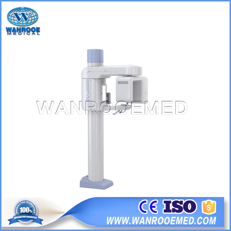

Oral and maxillofacial CBCT radiography system is equipped with 12 cm x 12 cm flat panel detector, which can be widely used in oral and maxillofacial surgery, orthodontics, orthognathic surgery, dental implant, endodontics and arthroscopic surgery.
PLX3000A CBCT Radiography System X-Ray Machine Features :
1. Adopts state-of-art flat panel detector of high resolution, slight anamorphose and uniform brightness.
2. Adopts pulse exposure mode, X-ray source can be only activated when needed. During 18s rotating process, X-ray only lasts 4~8s, which effectively reduces checking dose, reaches international advanced level.
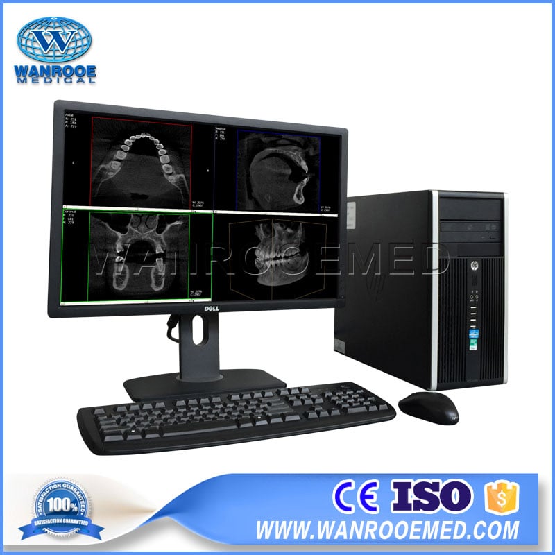
3. PLX3000’s exclusive 3D construction algorithm converts primitive 2D sequence projection to 3D volume image and offers highly clear sectional image in any angle and any position.
4. Highly clear oral panoramic image can be extracted through 3D volume image.
5. Image can be used to produce report, print or save report file.
6. Multiple radiography modes can be offered including standing, seating or wheelchair posture.
I. Technical parameters
1. Electric parameters
1.1 Anode voltage: 60~92 KV
1.2 Anode current: 1~15 mA
1.3 Max. output power: 1.38 KW
1.4 Parameter adjustment range: 60~92 KV, 1~15 mA
1.5 Information display: KV, mA, information of up/down position, characteristics of human body, laser designation and locating
2. X-ray tube assembly
2.1 Tube model: D-054SB (Toshiba)
2.2 Focal spot: 0.5mm
2.3 Thermal capacity: 35KJ (50KHU)
2.4 Radiography mode: CBCT
3. Imaging system (THALES flat panel detector 650HD-E)
3.1 Image receiving size: 12cm X 15cm
3.2 Pixel size: 150 um
3.3 Valid field of view: 80 (diameter) X 90 (height)
3.4 Display resolution: 1280 x 1024
3.5 Rotary angle: 200°
3.6 Rotary time: 18 S
3.7 Exposure time: 4~8 S
3.8 Scan number: 400 frames
3.9 Reconstruction time: ≤18 S
3.10 Voxel size: 0.2 mm
3.11 A/D conversion: 14 bits
3.12 Acquisition mode: pulse mode
3.13 Detector type: CMOS
3.14 Pixel matrix: 960 x 786
3.15 Resolution: 2D ≥ 3.1 lp/mm
4. Structural performance
4.1 Stand column up/down range: 1000 mm
4.2 Noise:<70dB
4.3 Anti-collision protection: YES
4.4 Complete machine weight: 260 Kg
4.5 Dimension: 788 mm (W) x 1090 mm (L) x 2200 mm (H)
4.6 Power requirement: 220 V/50 Hz/2 kVA
4.7 Space requirement: 5 sqm, single side 2 m
4.8 Radiology room should be of at least 2 mm lead equivalent
4.9 Electrical control device size: 340 mm (W) x 550 mm (D) x 650 mm (H)
II. Standard configuration
1. Rack 1 set
2. High frequency generator 1 set
3. 19” color general LCD, 24” color general LCD or
21” medical grade LCD (choose 1) 1 pc
4. 12 cm x 15 cm flat panel detector 1 set
5. Digital acquisition workstation 1 set
6. Electrical adjustable multi leaf collimator 1 set
7. Graphical LCD touch screen 1 pc
8. Wall mounted control box 1 pc
9. Electrical control cabinet 1 unit
10. Printer (Optional) 1 unit
Strive to Improve Medical Conditions, Reduce Health Care Costs
© 2018-2020 Jiangsu Rooe Medical Technology Co., Ltd. All rights reserved. Site Map